
Equine Complex Vertebral Malformation (ECVM) — Part 4: Diagnosis, Imaging, and Clinical Challenges
- Dr. Beth Byles, DVM

- Sep 8, 2025
- 1 min read
Diagnosing ECVM begins with a thorough clinical examination. Veterinarians assess posture, neck flexibility, and movement, watching for asymmetry, shortened stride, or resistance to bending—often early indicators of cervical involvement.
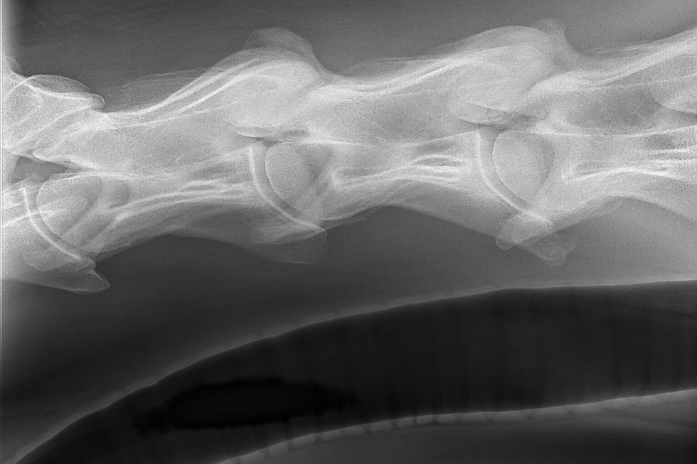
Radiographs are typically the first imaging step, focusing on C6, C7, and T1. Veterinarians evaluate bone shape, symmetry, and attachment points, looking for missing structures, fused elements, or rib transpositions. Imaging this region can be technically difficult due to the horse’s size and heavy musculature, and multiple views are often required. Larger horses may need referral to facilities with high-powered imaging systems.
Advanced imaging, particularly CT, provides detailed three-dimensional views of the cervical spine and is excellent for characterizing complex malformations. However, access remains limited. Many equine hospitals lack CT units large enough for the neck, and scans typically require general anesthesia, which carries inherent risks.
These diagnostic challenges mean ECVM may be underdiagnosed. A combination of clinical findings, imaging, and collaboration between veterinary professionals is often necessary to reach an accurate conclusion.
Next: practical strategies for managing horses diagnosed with ECVM.


Comments